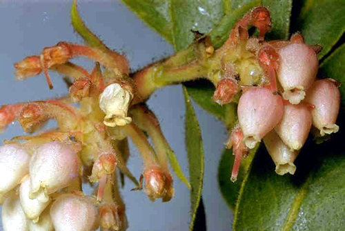
Arbutoid mycorrhizasArbutoid mycorrhizas are, like those of Ericoid and Monotropoid mycorrhizas, found in the plant order Ericales. Like ericoid mycorrhizas, the family Ericaceae is represented, with arbutoid mycorrhizas being formed in the genera Arctostaphylos and Arbutus (Fig. 1) . Arbutoid associations are also found in the Pyrolaceae family of the Order Ericales.
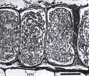
The fungi of arbutoid mycorrhizas are basidiomycetes, often the same fungal species that form ectomycorrhizal associations. Indeed, the structures of these two mycorrhizal types are very similar, and the assumption is that they operate similarly as well. In any given area of forest therefore, it is the particular taxon of plant which determines the type of mycorrhiza formed. Root structures of plants in the Arbustus and Arctostaphylos genera are differentiated into long and shoot roots, similar to those of forest trees. A fungal sheath or mantle of between 20 and 80 mm covers the roots, as in ectomycorrhizas. Nutrients scavenged by the mycelium and rhizomorphs in the soil have to pass through the sheath and into the short roots. The sheath can also provide an important store of nutrients, to be released to the plant when nutrient levels are sufficiently depleted. Also similar to ectomycorrhizas, arbutoid associations produce an intercellular Hartig net, usually restricted to the outer layer of root cells (Fig. 2). This 'paraepidermal' net is also seen in ectomycorrhizas in the majority of angiosperms. A hypodermis, formed by deposits of suberin and a Casparian strip in the outer layer of cortical cells, prevents deeper penetration of the Hartig net.
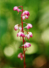
A major difference between the arbutoid and ectomycorrhizal association is that the hyphae of the former do actually penetrate the outer cortical cells, and fill them with coils, which the hyphae of ectomycorrhizas do not. The intracellular coils, along with the mantle sheath and Hartig net are the diagnostic features of arbutoid mycorrhizas. Root systems of species of Pyrola (Fig. 3) do not resemble those of ectomycorrhizas as much as do Arbustus and Arctostaphylos. All species of Pyrola, with the exception of P. uniflora, produce an extensive system of white rhizomes, with narrow adventitious roots. Colonisation by fungi increases the diameter of Pyrola roots, and the amount of branching also rises. The tips of colonised roots are usually transparent, turning brown or black with age.
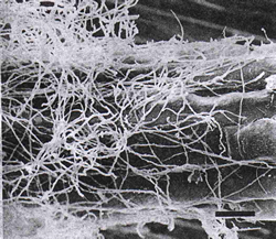
Colonisation occurs a few millimetres behind the root tip, where hyphae form a surface weft (Fig. 4). A full sheath is not formed. Hyphae penetrate intercellularly, forming a Hartig net, again of the paraepidermal type. Hyphae also penetrate intracellularly, growing extensively within the plant cells. |
To skip to other mycorrhizal types, click below: |
|||
Close the window to return to your previous page |
|||