
12.10 Mushroom mechanics
It is obvious that co-ordinated inflation plays a central role in differentiation of the cells and tissues which make up the fruit body, but it seems likely that cell inflation has a profound bearing on the morphogenesis of the fruit body as a whole. Again, our examples are concentrated on the ink cap mushroom Coprinopsis and its relatives, but they exemplify general points of structure that apply equally to other fungi.
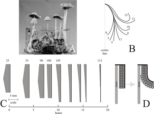
Except for the descriptions given by Buller (1924, 1931), discussion of cap morphogenesis in ink caps is generally restricted to the role of autolysis (the self-digestion of the gill tissue that produces the ‘ink’) in the removal of spent gill tissue which might otherwise interfere with spore-fall from the fruit body. However, during this autodigestive period the cap passes through a profound change in shape before autolysis: the initially vertical orientation of the gills is transformed to a horizontal one (Buller, 1924, 1931). In Coprinopsis cinerea this process is completed in about 6 h and is achieved by a gradual rolling-back of the edge of the cap, which, together with radial splitting of cap and gill tissues, allows the cap to open like an umbrella. The mechanical process that achieves this emerges from measurement of the profile contour length of the cap at successive stages during expansion, which shows that it remains constant in the final autodigestive phase (Fig. 24). This is true even though:
- as paraphyses differentiate their inflation must increase the area of the gill, and
- although spent gill tissue is removed by autodigestion a considerable portion remains intact until very late in cap expansion.
So, a gill that is expanding in area is bounded by a flexible but evidently inextensible band of cap tissue, a combination which will inevitably lead to an outward curvature (Fig. 24). Cap tissue becomes the most important structural members after the autodigestive process has removed the bulk of the gill, but initially paraphyseal inflation provides the major expansionary force and structural integrity (Ewaze et al., 1978; Moore et al., 1979).
 |
|---|
| Fig. 24. Final maturation of the fruit body cap of Coprinopsis cinerea. Left to right: A. Photograph of the organism fruiting on compost in a 9 cm diameter crystallising dish. B. Diagrams recording the way in which the profile of the cap changes during its final phase of expansion. Consecutive photographs were made of the same fruit body as it matured and tracings were then made of the upper edge of the cap profile. Number show hours elapsed since observation started at time zero. During the course of the sequence shown the stem extended from 25–93 mm, spore discharge commenced about 5 h from the start of observation. C. Scale drawings of gill growth and autolysis. At regular intervals in the final stages of fruit body development a small segment was surgically removed from an otherwise undisturbed fruit body and preserved in formalin. Subsequently, the outer contour length (length of cap flesh or pileipellis) was measured and the depth of gill tissue measured at the top, centre and bottom of a suitable primary gill in each segment. In these diagrams the shape of the cap is ignored (for which see B); the vertical bars represent the contour length and the shaded area represents the gill lamella. The numbers above the diagrams record the length (in mm) of the stem at the time the sample was taken. In this specimen, spore discharge began at about the 7th hour. These observations show that the cap flesh remains intact and extended throughout the entire period of gill autolysis, and that for the final period of cap development the length of the cap flesh is unchanged. D. Development of the fruit body cap in Coprinopsis cinerea. Diagram to show how cell expansion in the hymenium can account for the outward bending of the gills (as indicated in the profiles shown in B) if it is assumed that the cap flesh (pileipellis) acts as a flexible but inextensible outer boundary (shown as a thick black line). Diagrams redrawn after Moore et al. (1979). |
We have now brought attention to several structural characteristics of the Coprinopsis cinerea fruit body that are the result of mechanical processes:
- the regular ‘parallel’ arrangement of gills (CLICK HERE for a reminder);
- conversion of the stem to a hollow cylinder (CLICK HERE for a reminder);
- and now the final morphogenesis occurring during maturation of the fruit body cap.
It is an important general point that several distinctive aspects of mature multicellular structures in fungi are the result of mechanical interactions between tissues that are inflating to different extents. They result in a very exact, and recognisable, final morphology; but that final morphology is not explicitly specified in the genome. Rather, the genome specifies a programme of metabolic and structural activities which, if played out correctly will result in that characteristic morphology.
Updated July, 2019